Skin cancer around the eye: how it is removed
Two patches at the inner corners of a lady's eyes had been treated as eczema for years. Both were basal cell carcinomas. What periocular skin cancer actually looks like, why it is so easily missed, and why the method of removal matters more than the operation itself.
A lady in her seventies was referred to me with what had been assumed to be a persistent patch of eczema at the inner corner of her eye. It had been there a long time, crusting and bleeding occasionally, never quite healing. There was a similar patch at the inner corner of the other eye.
Both were basal cell carcinomas.
This is the typical story. Skin cancer around the eye rarely announces itself. It can be a bump or a lump, as you might expect a tumour to be. But just as often it is a sore that will not heal, a persistent red patch, a small pearly nodule, or an area where the eyelashes have quietly disappeared. Patients assume it is irritation, blepharitis or dry skin, and a good number of clinicians assume the same. Many of the ones I see have been present for months to years before anyone reaches a diagnosis.
The good news is that these tumours are almost always curable. The part that matters is how they are removed.
What these tumours actually look like
The photographs on this page are all of one patient, published with her consent. She has been under my care for several years and has had three separate basal cell carcinomas around her eyes. All three were photographed before surgery, and between them they cover most of the reasons these tumours get missed.
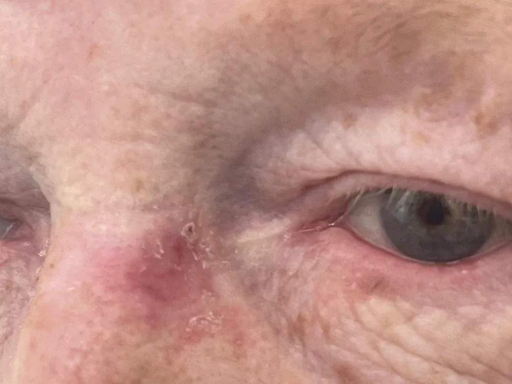
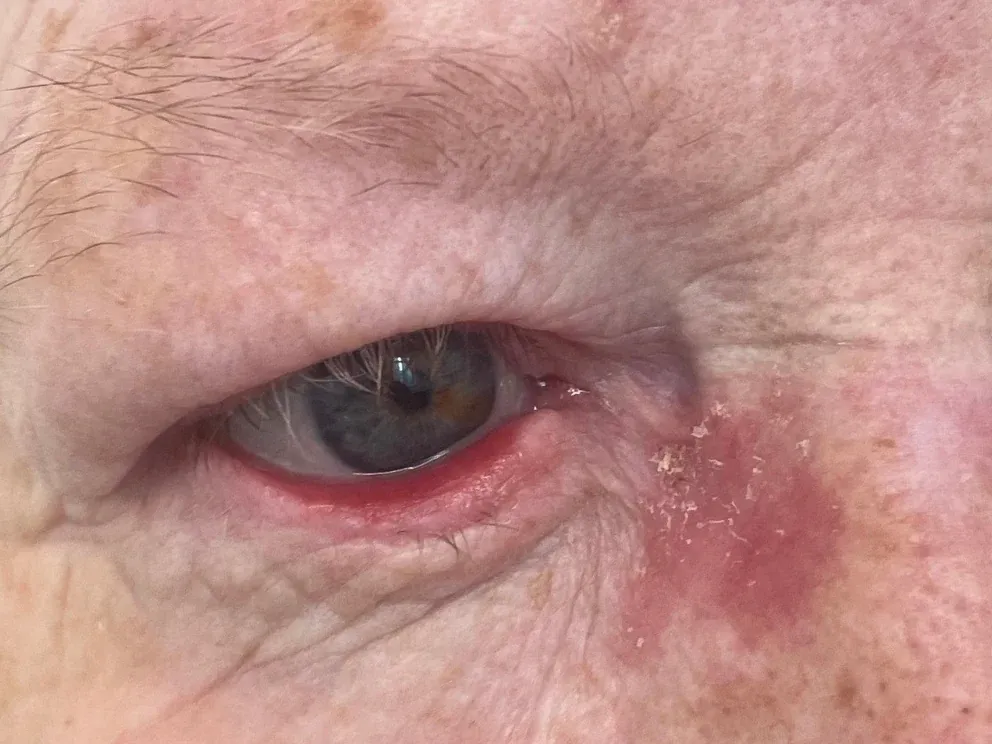
Both of those are cancer. They look like eczema: red, faintly scaly, mildly crusted, entirely unremarkable. Nothing about either one announces malignancy, and a great many are treated with steroid cream or emollient for months before anybody becomes suspicious.
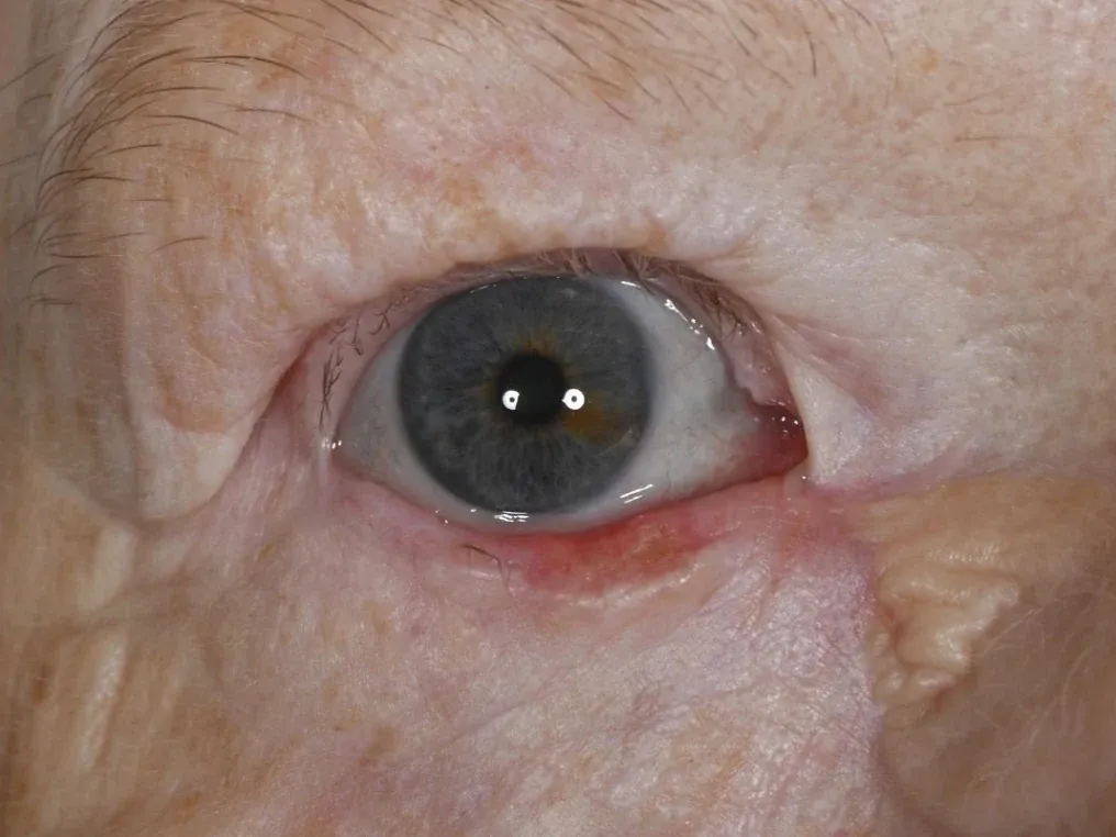
The third tumour, on the lower eyelid margin, is the other commonly missed presentation. Redness and irritation along the lid margin is the appearance most often labelled as blepharitis and treated with lid hygiene, sometimes for a year or more.
The features that should raise suspicion are not dramatic:
- a lesion or patch present for months that has never fully healed
- repeated crusting or bleeding, usually described as “it scabs over and then comes back”
- loss of eyelashes in one area
- distortion or notching of the eyelid margin
- redness or irritation confined to one eye, or to one part of one eyelid, when the other side is normal
- a pearly, raised edge, sometimes with fine visible blood vessels
- a chalazion that keeps returning to exactly the same spot
Two of those deserve emphasis for clinicians as much as for patients. One-sided blepharitis is not blepharitis until proven otherwise, and a chalazion that recurs in the same place deserves a biopsy rather than a third incision.
What these tumours are
Somewhere between 5 and 10 percent of all skin cancers occur in the region around the eye, and the overwhelming majority of those, roughly 90 to 95 percent, are basal cell carcinomas. They grow slowly and essentially never spread to other parts of the body. What they do instead is grow locally, invading whatever is next to them, which around the eye means the eyelid, the tear drainage system, and in advanced cases the eye socket itself.
Squamous cell carcinoma is less common but more aggressive. It can spread, and left untreated it can shorten life. Rarer still are melanoma and sebaceous gland carcinoma, which can behave more aggressively again.
Location matters. Around half of periocular basal cell carcinomas affect the lower eyelid. The medial canthus, the inner corner where the eyelids meet the nose, is the next most common site and the most anatomically demanding, because the tear drainage system runs through it and the tissue planes there are complex.
Why the eye is different
Skin cancer on the back or the arm can be removed with a generous margin of normal tissue and very little consequence. Around the eye, every millimetre of tissue is doing a job. The eyelid protects the eye, spreads the tear film with every blink, and drains tears into the nose. Take too much and you create a functional problem: an eyelid that will not close, a lid that turns outward, lashes that rub, an eye that waters permanently.
The eyes are also where other people look. They carry most of facial expression, and they are one of the hardest parts of the body to hide. Patients are right to care how the result looks.
So there are two competing pressures. Remove enough to be certain the cancer is gone. Remove as little as possible to preserve function and appearance. Every technique below is an attempt to resolve that tension.
Option one: wide local excision
The tumour is removed with a predetermined margin of normal-looking tissue around it, typically three to four millimetres, and the defect is reconstructed immediately in the same operation. The tissue goes to the laboratory afterwards and the result comes back a week or two later, confirming whether the margins were clear.
When it is appropriate. Small, well-defined, previously untreated tumours in a favourable location, where the edges are clearly visible, the repair is straightforward, and the histology is likely to be a simple nodular basal cell carcinoma.
The advantage. One operation, one anaesthetic, immediate reconstruction, and the whole thing is finished sooner.
The disadvantage. You find out whether it worked afterwards. If the margins come back involved, the reconstruction has already been done, and further surgery means operating through a healing repair. Published recurrence rates for periocular basal cell carcinoma after wide local excision sit at around 6 percent, roughly double the rate for margin-controlled techniques.
That difference is the entire reason the other approaches exist.
Option two: Mohs micrographic surgery
Traditionally the gold standard for high-risk facial skin cancer. The tumour is removed in thin layers, and each layer is processed and examined under the microscope immediately, while the patient waits. The surgeon maps exactly where any remaining tumour is and takes a further layer from that point alone. The process repeats until the margins are clear.
The elegance of it is that, done properly, it removes the minimum possible tissue while examining effectively the whole margin rather than sampling it. Published recurrence rates for periocular basal cell carcinoma after Mohs are around 3 percent.
We offer Mohs both on the NHS and privately. The excision is carried out by our local Mohs surgeons and I perform the eyelid reconstruction shortly afterwards. We work closely together and meet monthly to discuss all of our joint patients, which means the repair is planned with knowledge of exactly what was taken and why.
The practical constraint is capacity rather than availability. Mohs needs a specialist surgeon and a laboratory processing frozen sections in real time, which limits how many cases fit into a session, so waiting times are often longer than for other methods of removal. For a slow-growing nodular basal cell carcinoma that rarely matters. For a more aggressive tumour, or one already threatening the eyelid margin or the tear drainage system, waiting is itself a cost.
Option three: slow Mohs
Slow Mohs achieves the same margin control using conventionally processed tissue rather than frozen sections. The difference is timing, and often a shorter wait to get started.
Here is how it works in our service.
- Stage one. The tumour is marked out and removed with a narrow margin of one to two millimetres of normal-looking tissue. A further 1 mm margin is taken from around the edges of the excision and examined by the pathologist in its entirety. It is orientated and marked, so that if tumour is still reaching the edge of the specimen in one place, or in several, I know exactly where to take more from.
- The wait. The tissue is processed conventionally, fixed and embedded in paraffin, then examined. This takes a few days rather than an hour, but the sections are of higher quality than frozen sections, which matters for interpretation.
- Stage two, if it is needed. If tumour is present at a particular edge, I take a further layer from precisely that point and nowhere else. This repeats until the margins are clear.
- Reconstruction. Once clearance is confirmed, the defect is repaired.
In our own service evaluation at Portsmouth, following patients for five to seven years, half the tumours were cleared in a single stage and 95 percent within two. Only two patients needed more than two stages. Recurrence at five years occurred in one patient, a rate of 2.4 percent, which sits comfortably within the range published for conventional Mohs. Our findings are consistent with a similar series from Newcastle.
The honest trade-off is that this means more than one visit and a period with an open, dressed wound. The wound is covered, comfortable and looked after, and most patients find the whole thing less troubling than they expected once it has been explained. But it does need explaining properly rather than glossing over it.
Which approach for which patient
The decision depends on the tumour, not on preference.
Margin-controlled excision, whether conventional or slow Mohs, is preferred for tumours at the medial canthus, tumours involving the eyelid margin, recurrent tumours, tumours with indistinct clinical edges, aggressive histological subtypes such as infiltrative or morphoeic basal cell carcinoma, large tumours, and squamous cell or sebaceous carcinomas.
Wide local excision is reasonable for small, well-defined, previously untreated nodular basal cell carcinomas away from the lid margin and the medial canthus.
A significant proportion of the tumours I see fall into the first group, which is why margin control is my default in this region. The consequence of getting it wrong here is not simply a recurrence, but a recurrence in scarred tissue where the anatomy has already been altered and further surgery is harder.
Choosing between Mohs and slow Mohs is usually a practical decision rather than an oncological one, since the recurrence rates are comparable. Conventional Mohs completes the excision in a single visit, which some patients much prefer. Slow Mohs can usually be started sooner and produces higher-quality sections, at the cost of a staged procedure. Where a tumour is behaving aggressively or already involves the lid margin, the shorter wait to treatment can be the deciding factor. I discuss both, and the right answer differs from patient to patient.
What about non-surgical treatments?
Creams, cryotherapy and photodynamic therapy have a place for superficial basal cell carcinomas elsewhere on the body. Around the eye they are used cautiously, because the margins cannot be checked, the surface of the eye can be damaged by the agents used, and a recurrence hidden underneath a healed surface is difficult to detect until it is advanced. Radiotherapy remains an option for patients unsuitable for surgery, though it carries its own consequences for the eye and the tear film.
After treatment
Most patients are followed for several years afterwards. Having had one skin cancer is the strongest predictor of having another, so surveillance is about the rest of the face as much as the treated site. Sun protection genuinely matters after treatment, not only before.
The patient whose photographs appear above illustrates the point well. She developed three separate basal cell carcinomas around her eyes over about two and a half years, each identified and treated as it arose. None of them was a recurrence of the last. They were new tumours on skin that had already declared its susceptibility.
The one recurrence in our five-year series appeared at 48 months, which is a further reason follow-up extends well beyond the first year or two.
Getting seen
Any lesion around the eye that has been present for more than a few weeks and is not healing, particularly one that bleeds or crusts repeatedly, that has caused eyelash loss, or that distorts the lid margin, should be assessed. Persistent one-sided blepharitis and a chalazion that keeps coming back in the same spot also deserve a proper look, because those are the presentations most often missed.
Diagnosis is usually clinical, sometimes confirmed with a small biopsy first. Being seen early means a smaller tumour, a smaller defect, a simpler reconstruction and a better result.
Continue reading. Removing the tumour is half of the problem. What happens next depends entirely on what the tumour took away: rebuilding the eyelid after skin cancer surgery follows the same patient through three different reconstructions, and shows what they look like at three months and at two years.
Procedure pages this note touches on.
A persistent eyelid lump, cyst or stye that has not settled with conservative measures. Most are benign and easily removed in the clinic or theatre.
A two-stage approach to eyelid skin cancer: careful excision with margin control, followed by reconstruction tailored to the size and site of the defect.
Other recent notes.
Is eyelid surgery funded on the NHS?
There is no national rule. Whether an eyelid operation is paid for depends on which operation it is and which integrated care board your postcode sits in, and neighbouring boards reach opposite answers on the same patient. Here is how to find out where you stand.
Rebuilding the eyelid after skin cancer surgery
One patient, three basal cell carcinomas, three entirely different repairs. What determines the reconstruction, why a large lower lid defect means a closed eye for a month, and what a graft looks like at three months compared with two years on the same face.