Rebuilding the eyelid after skin cancer surgery
One patient, three basal cell carcinomas, three entirely different repairs. What determines the reconstruction, why a large lower lid defect means a closed eye for a month, and what a graft looks like at three months compared with two years on the same face.
The lady whose tumours appear in skin cancer around the eye: how it is removed has been under my care for several years. She has had three basal cell carcinomas: one at the inner corner of each eye, and a third on her right lower eyelid. They did not arrive together. Each was treated as it appeared, over about two and a half years.
All three were removed with margin control. All three were reconstructed differently. The two inner corners were repaired with skin grafts taken from in front of the ear. The lower eyelid needed a two-stage operation, borrowing tissue from the upper lid to rebuild the inner lining, with a skin graft for the outer layer.
Three tumours in one patient, three different repairs.
That is the honest answer to the question almost everybody asks, which is “what will you do to fix it?” There is no standard reconstruction. What gets rebuilt is determined by what the tumour took away.
Her photographs appear throughout this page with her consent, and they are useful for a second reason. Because her operations were spread over years, you can see a two-year-old graft and a three-month-old reconstruction on the same face. That comparison is the most honest illustration of healing I can offer.
First, what the eyelid is made of
Everything about reconstruction follows from this, so it is worth thirty seconds.
The eyelid is built in two layers, like a sandwich.
- The front layer is skin, and the thin muscle that closes the eye. Eyelid skin is the thinnest on the body.
- The back layer is a firm plate that gives the lid its shape, lined by a smooth, moist membrane that sits directly against the surface of the eye.
Both matter, for different reasons. The front layer determines how the lid looks and whether it can close. The back layer determines whether it holds its shape, and whether the surface against the eye is smooth. A rough or bulky inner lining will damage the cornea with every blink, which is why that layer cannot be improvised.
The question that determines everything
When a tumour is removed, what is left is a defect, and the reconstruction is decided by asking one question: which layers are missing, and how much of them?
There are three broad answers, and they lead in quite different directions.
Answer one: only the front layer is missing
This is the commonest situation. It is what happens with most tumours at the inner corner of the eye, and with tumours on eyelid skin away from the margin. The firm plate and its lining are untouched. Only skin needs replacing.
Several options, roughly in order of increasing complexity:
- Let it heal on its own. Small defects in concave areas, particularly the inner corner, often heal beautifully if they are simply dressed and left. This is not a shortcut. In a hollow it frequently gives a better contour than a graft, which can sit slightly proud. The trade-offs are a longer healing period with a wound to dress, and a little less predictability, since healing tissue can tighten and pull.
- Close it directly. If the defect is small and the skin around it is mobile, the edges can be brought together and stitched. Ageing skin is more forgiving here, which is one of the few advantages of presenting later in life.
- Move nearby skin in, as a flap. Skin from immediately next to the defect is slid, rotated or advanced into the gap while staying attached to its own blood supply. The match is excellent, because the tissue comes from millimetres away, and the design follows the natural creases of the face so the scars fall into lines that already exist. Adjacent skin is not always available, and in this patient it was not, because of the other tumours nearby.
- Bring skin from elsewhere, as a graft. A piece of skin is taken from another site, thinned, and stitched in, surviving on nourishment from the tissue beneath it. This is what was done at both inner corners here.
Where graft skin comes from matters more than people expect. The donor site is chosen to match the thinness, colour and texture of eyelid skin. In practice that means the upper eyelid of one or both eyes, which is the closest match and occasionally a small bonus where there is excess skin there anyway, or the skin in front of or behind the ear, and sometimes above the collarbone or the upper arm. This patient had a large defect to fill on both sides and not enough spare skin in her upper lids, so I used skin from in front of each ear: it matches well, the supply is reliable, and the donor scar hides in a natural crease.
The same two corners, before and after
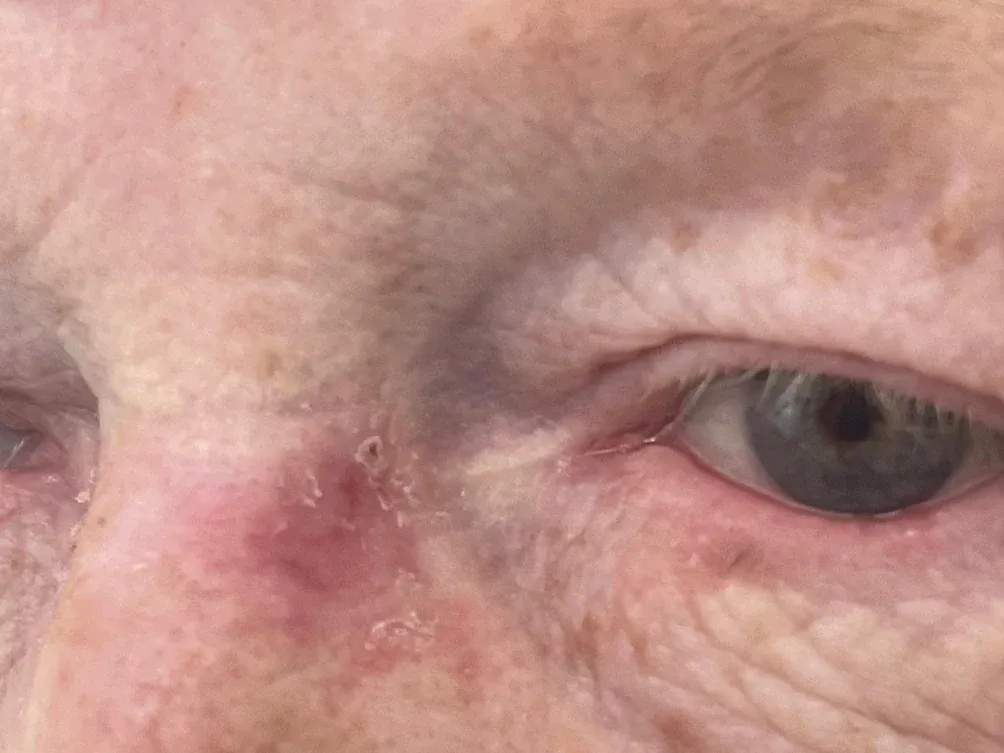
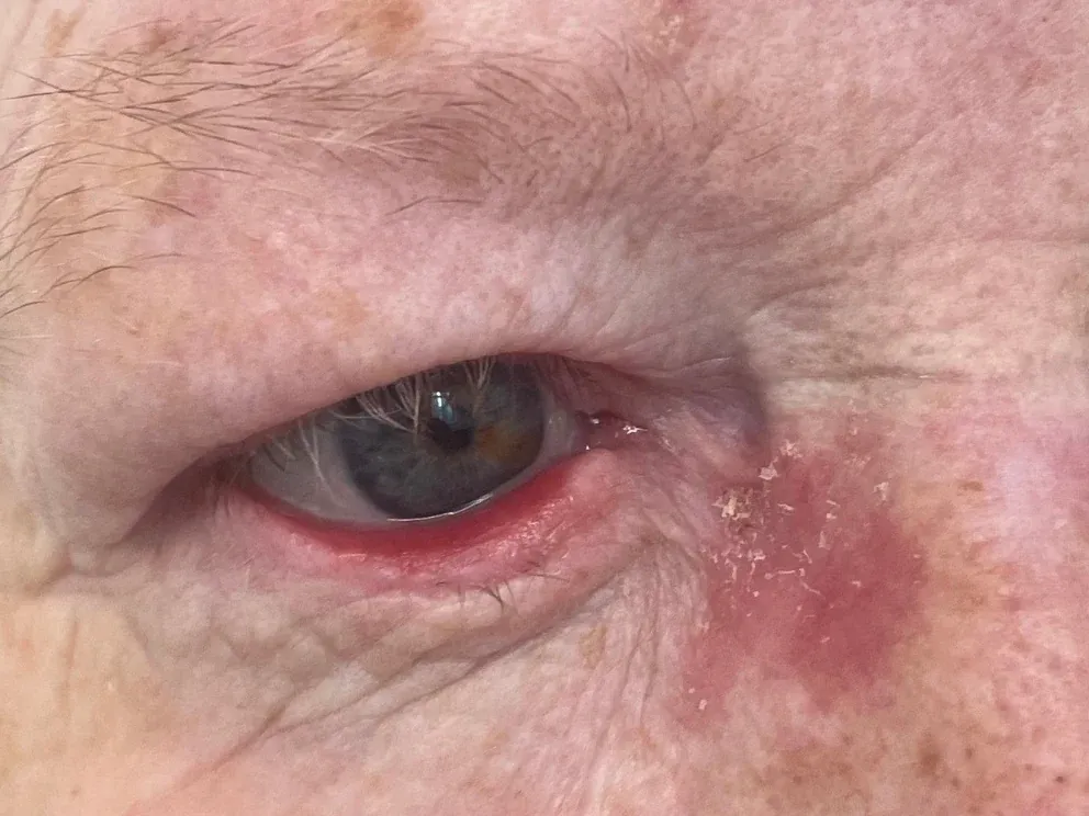
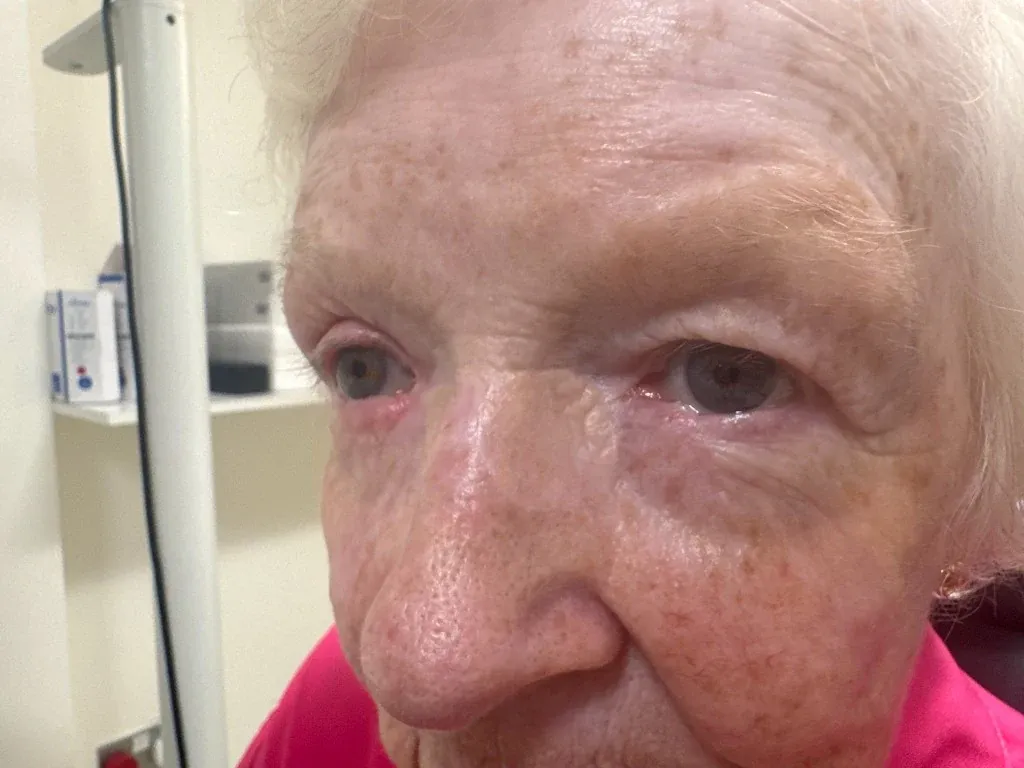
Those are the same two inner corners, first as tumours and then at two years and two and a half years after grafting. The grafts are pale, thin, and difficult to find. That is what a settled graft looks like, and it is worth knowing that it takes this long to get there.
Answer two: the whole thickness is missing, but only a small amount
Where a tumour has been taken through the full thickness of the eyelid, both layers need rebuilding. If the defect is small, the eyelid can often be rebuilt with itself.
The cut edges are brought together and the lid margin carefully aligned, sometimes with a small release at the outer corner to gain a few millimetres of slack. Up to about a quarter to a third of the eyelid can usually be closed this way, and more in older patients whose lids are naturally looser.
This is the best outcome available, because every layer is rebuilt with the correct tissue. Where it is possible, it is what I do.
Answer three: the whole thickness is missing, and a lot of it
This is the demanding situation, and it is where the two-layer principle becomes a hard rule.
Both layers cannot be rebuilt with grafts. A graft has no blood supply of its own and depends entirely on the tissue it is laid against. Put a graft on a graft and neither survives. So when a large full-thickness defect is rebuilt, at least one of the two layers must be a flap: tissue moved from nearby while staying connected to its own circulation.
That single constraint explains most of what follows, and it is the reason large lower lid reconstructions are more elaborate than patients expect.
The Hughes flap
For substantial full-thickness lower eyelid defects, one of the most reliable answers is a two-stage operation called a Hughes tarsoconjunctival flap.
- Stage one. A flap of the firm plate and its moist lining is raised from inside the upper eyelid and brought down into the lower lid defect, rebuilding the back layer. Because the flap has to stay attached to its blood supply in the upper lid, it forms a bridge across the eye. The front layer is then rebuilt over it, usually with a skin graft, often from the upper eyelid. The consequence is that the eye on that side is temporarily closed by the bridge of tissue.
- The wait. Usually two to four weeks, during which the transferred tissue develops its own blood supply from the lower lid.
- Stage two. The bridge is divided, releasing the eyelids, and the new lid margin is contoured. This is a much smaller procedure under local anaesthetic.
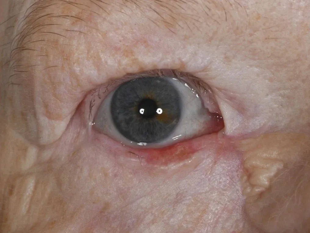
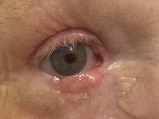
Why accept a closed eye for a month? Because for these defects it produces the most reliable and predictable result available. The rebuilt lid has proper structure, a smooth lining against the eye, and a stable margin that holds its position over years.
It is worth being plain about the inconvenience. Having one eye closed for several weeks is a genuine imposition, and depth perception is affected in a way that matters for driving and for stairs. Patients whose other eye sees poorly may not be suitable at all, and I ask about this specifically, because temporarily closing somebody’s better-seeing eye is a serious decision rather than a technical detail. Most people manage well when they know in advance what they are agreeing to.
Similar principles apply to large upper lid defects, though the reconstruction differs, because the upper lid does most of the work of closing the eye and is less forgiving.
The tear drainage system
Tumours at the inner corner sometimes involve the channels that drain tears into the nose. Where those are lost, the eye may water persistently afterwards.
This can usually be dealt with at the time of the repair, often with a temporary stent left in place while the tissues heal. Where a watering eye persists, the later options include creating a new drainage pathway into the nose, sometimes with a small glass bypass tube if the original channels have been destroyed.
What to expect afterwards
Some honest expectations, because this is where most of the anxiety sits.
Grafts look worse before they look better, and the difference is measured in years rather than weeks. The photographs on this page make the point better than any description can. The grafts at both inner corners are two and two and a half years old: pale, thin, and blended into the surrounding skin, so that you have to know where to look to find them. The right lower eyelid reconstruction is three months old, still pink and a little thickened, and still remodelling. Both belong to the same person, photographed on the same day. The three-month appearance is not a worse result than the two-year one. It is the same result, earlier.
In the first weeks and months a graft or a flap is pink, sometimes red or purple, often crusted at the edges, and can feel firm. It looks like a visible patch, and almost every patient worries at this stage that something has gone wrong. Over roughly a year the colour fades towards the surrounding skin and the firmness softens, and it goes on improving quietly for a good while after that.
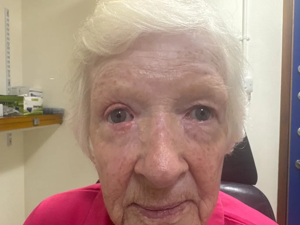
Notice how little of it registers at normal conversational distance. Patients examine their own reconstruction from six inches away in a bathroom mirror. Nobody else ever sees it that way.
Timelines. Bruising and swelling settle over two to three weeks. Appearance improves markedly between one and three months. Grafts continue to fade and soften for a year and beyond, and the two-year results on this page are noticeably better than they were at twelve months. Judging the final result before six months is judging it far too early, and that catches almost everyone out.
There will be a scar. Around the eye scars settle exceptionally well, because the skin is thin and the blood supply is generous. The aim is a scar that is hard to notice, not no scar at all.
Function comes before appearance. The priorities, in order, are complete removal of the cancer, a lid that closes and protects the eye, a lid that sits in the right position, a lid that drains tears effectively, and then appearance. Usually these align. Where they conflict, that is the order in which I decide.
Small revisions are normal, not failures. A minor adjustment under local anaesthetic some months later, refining a lid margin or thinning a graft, is a routine part of the process.
The commonest imperfections are a small notch in the lid margin, a lid that turns slightly outward as scar tissue contracts, and a graft that stays a little different in colour. In our own five-year series, 14 percent of patients developed some degree of ectropion, one of whom needed a repair.
Why this belongs with an oculoplastic surgeon
Removing a skin cancer and rebuilding an eyelid are different skills, and around the eye they have to be planned together. Our Mohs surgeons carry out the excision and I carry out the reconstruction, and we meet monthly to discuss all of our joint patients, so the repair is planned with full knowledge of what was taken and why.
An oculoplastic surgeon is an ophthalmologist first, with subspecialty training in eyelid and orbital surgery. That matters because both the excision and the repair are then planned by somebody who knows what the eyelid has to do, not only how it should look. The lining against the eye must be smooth, or the cornea is damaged with every blink. Lash direction matters. The tear drainage system runs directly through the area where these tumours most often occur. And an eyelid that looks acceptable but does not close properly is a failed reconstruction, however good the photograph.
The reassuring part
The great majority of patients come through this with an eye that works normally and an appearance that draws no attention. The interim is the hard part. Knowing in advance that grafts stay pink for months, that a Hughes flap means a closed eye for a while, and that three months is not the final result makes that interim considerably easier.
Start at the beginning. Skin cancer around the eye: how it is removed covers what these tumours look like, why they are so often mistaken for eczema or blepharitis, and the difference between wide local excision, Mohs and slow Mohs.
Procedure pages this note touches on.
When the lower eyelid sags away from the surface of the eye, leaving it red, sore and watering. Surgery restores the normal eyelid position.
A two-stage approach to eyelid skin cancer: careful excision with margin control, followed by reconstruction tailored to the size and site of the defect.
A structured assessment of the watering eye, to identify the cause precisely before recommending any treatment.
Other recent notes.
Is eyelid surgery funded on the NHS?
There is no national rule. Whether an eyelid operation is paid for depends on which operation it is and which integrated care board your postcode sits in, and neighbouring boards reach opposite answers on the same patient. Here is how to find out where you stand.
When the tear duct is partly blocked: which operation, and does it last?
A syringing test that passes proves the pipe is not sealed shut. It does not prove it works. Why partial and functional tear duct obstruction is so often dismissed, what the two operations for it are, and why they look identical at three months and very different at eight years.
Watery, sticky eyes in babies and toddlers
A blocked tear duct is one of the commonest things to affect a newborn, and most open on their own without treatment. What helps in the meantime, why repeated antibiotic drops are rarely the answer, when an operation is worth considering, and why I do that operation under endoscopic guidance.