Watery, sticky eyes in babies and toddlers
A blocked tear duct is one of the commonest things to affect a newborn, and most open on their own without treatment. What helps in the meantime, why repeated antibiotic drops are rarely the answer, when an operation is worth considering, and why I do that operation under endoscopic guidance.
I saw a little girl in clinic this week, a few days short of her first birthday, whose eyes had been watering and sticky on both sides since she was born. At around six months the left eye cleared up by itself, shortly after a cold. The right carried on.
On examination the right eye was streaming but otherwise entirely healthy: clear cornea, clear conjunctiva, no infection, no swelling of the tear sac, and her vision developing normally.
Her parents had two reasonable questions. Does she need an operation, and if so, when?
The honest answer to the first is that she may well not. Her left eye sorted itself out with nobody doing anything, which is exactly what usually happens, and there is a fair chance the right will follow. I will come back to the second question at the end.
What is happening
Tears are made continuously to keep the eye healthy, and they drain away through a small channel running from the inner corner of the eye down into the nose. That is why your nose runs when you cry.
In the last weeks before birth, the lower end of this channel opens into the nose as a thin membrane breaks down. In a good number of babies it has not quite finished the job by the time they are born. The channel is complete except for a membrane at the very bottom.
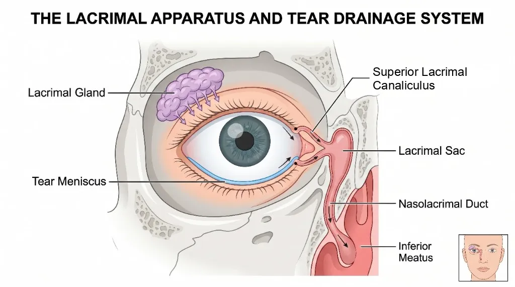
The tear drainage pathway. Tears drain from the inner corner of the eye through the canaliculi into the tear sac, then down the nasolacrimal duct into the nose. In a baby with a blocked tear duct, the hold-up is almost always right at the bottom of that pathway, where the duct opens into the nose.
Tears then have nowhere to go. They pool, spill over the lid, and run down the cheek. Because still tears are a good place for bacteria to sit, the eye becomes sticky, particularly overnight.
The medical name is congenital nasolacrimal duct obstruction. It affects up to one in five newborns to some degree, which makes it very common indeed. It is frequently bilateral, as it was in the child above, and the two sides do not necessarily resolve at the same time.
The reassuring part
Most of these open on their own. The membrane continues to break down after birth in the great majority of babies, and by the first birthday around nine in ten have resolved with no treatment whatsoever. Parents often notice it clear up shortly after a cold, as the duct opens.
Importantly, resolution does not stop at twelve months. A significant proportion continue to settle spontaneously beyond that age. This is why reaching the first birthday with a watery eye is not in itself a reason to operate, and why “let us wait a bit longer” is often the right advice rather than a fob-off.
What helps in the meantime:
- Cleaning. Cooled boiled water on cotton wool, wiping from the inner corner outwards. Stickiness is usually stagnant tears rather than infection, so it needs cleaning rather than treating.
- Massage. Gentle, firm downward pressure over the tear sac at the inner corner of the eye, beside the nose, several times a day. The aim is to generate a little pressure in the channel to help the membrane open. There is reasonable evidence it improves the chances of spontaneous resolution, and done gently it does no harm.
- Antibiotic drops only when genuinely needed. Repeated courses are one of the commonest patterns I see, and they usually treat the wrong thing. They clear a sticky eye briefly, but while the tears cannot drain, the stickiness returns. Occasional courses are reasonable for genuine infection. Repeated ones mean the underlying problem has not been addressed.
Seek help sooner if there is a red, swollen, tender lump at the inner corner of the eye, if your child is unwell, or if the eye is red and painful rather than simply wet and sticky. These are uncommon but need seeing promptly.
When treatment is worth considering
There is no fixed age at which waiting stops being reasonable. Historically intervention was recommended between one and two years, partly because recurrence appears higher when probing is done above the age of two. That figure needs interpreting carefully: the older group excludes all the children who had already resolved spontaneously and therefore never needed treating, so it is not comparing like with like.
In practice the decision depends on how much it is troubling the child and the family, whether both sides or one are affected, whether there have been episodes of infection, and how the eye looks. It is a conversation rather than a rule.
Where treatment is needed, the standard procedure is called syringing and probing. Under a short general anaesthetic, a fine probe is passed down the tear channel to open the membrane at the bottom, and fluid is passed through to confirm it drains freely into the nose.
It takes a few minutes. There is no cut, no scar and no stitches. Most children are home the same day and back to normal the next.
The part I do differently
Conventional syringing and probing is done blind. The surgeon passes the probe, feels it give way, flushes fluid through, and confirms drainage by feel and by seeing fluid appear. It works most of the time, and it remains the most commonly performed version of this operation in the UK.
I perform the procedure with a small endoscope passed into the nose, so I can see the lower end of the tear duct directly while I work. This matters for two reasons.
First, it confirms what has actually happened. Rather than inferring that the probe went where it should, I can see it emerge, see the membrane open, and see fluid drain.
Second, and more usefully, it shows what else is going on. A blocked tear duct is not always just a membrane. In our own series, most children having endoscopically guided probing needed something beyond a simple probe: the lower end of the channel opened up because it was too narrow, a bone in the nose gently moved aside because it was pressing on the duct, or a membrane divided under direct vision because a probe alone would not have cleared it. Some turned out to have inflammation in the nose contributing to their symptoms, which could then be treated properly.
Around four in five of the children we treated this way had either an additional surgical step or additional medical treatment that a blind procedure would not have identified.
Does it make a difference?
Yes, and by a meaningful margin.
Combining our results with those of another UK centre, giving nearly 250 procedures in total:
- Probing without endoscopy: around 80 percent success.
- Probing with endoscopy: around 90 percent success.
Roughly one child in ten is spared a second procedure by having the first done under direct vision.
For a parent the relevant currency is not percentages but general anaesthetics. Every failed procedure means another anaesthetic, another morning of a child who cannot eat beforehand, another wait, and usually a more involved second operation. Reducing how often that happens is the point.
What to expect on the day
- A short general anaesthetic, as a day case, home within a few hours.
- No cuts, no stitches, no dressing.
- A little blood-stained fluid from the nose or eye on the first day is normal.
- Antibiotic drops for a few days afterwards in most cases.
- Watering usually improves within days, though some residual watering while things settle is common.
- A review several weeks later to confirm it has worked.
If it does not work first time
A small proportion of children continue to water afterwards. The usual next step is repeat probing with a soft silicone tube left in the duct for two to three months to hold it open while it heals. Removing the tube needs a further brief anaesthetic, so this route means two additional general anaesthetics in total. This is worth knowing in advance, and is part of why getting the first procedure right matters.
A small number of children, usually those with an unusual anatomical cause, eventually need a more involved operation to create a new drainage pathway. This is uncommon in childhood.
The summary
A watery, sticky eye in a baby is common, is almost never dangerous, and usually resolves on its own. Repeated antibiotic drops are rarely the answer. Reaching the first birthday is not automatically a reason to operate, as many children settle after that.
Where it does persist and treatment is needed, a short procedure fixes it in the great majority of cases, and doing that procedure under endoscopic guidance improves the odds of getting it right first time.
Back to the little girl
I said I would return to the second question, which was when.
We decided to plan for surgery in a few months’ time. That gives her right eye a further opportunity to settle by itself, as her left one did, while making sure that if it does not, she is not waiting to start the process from scratch.
If it clears up before then, the procedure comes off the list and she never has the anaesthetic. If it does not, she has it done at a good age.
Figure: illustration licensed from Adobe Stock (2155135037). Adapted from the original, in which the upper channel was labelled the superior lacrimal duct; it is the superior lacrimal canaliculus.
Procedure pages this note touches on.
Scarless surgery for a blocked tear duct, performed entirely through the nose using an endoscope, with a high success rate and quick recovery.
A structured assessment of the watering eye, to identify the cause precisely before recommending any treatment.
Other recent notes.
Is eyelid surgery funded on the NHS?
There is no national rule. Whether an eyelid operation is paid for depends on which operation it is and which integrated care board your postcode sits in, and neighbouring boards reach opposite answers on the same patient. Here is how to find out where you stand.
When the tear duct is partly blocked: which operation, and does it last?
A syringing test that passes proves the pipe is not sealed shut. It does not prove it works. Why partial and functional tear duct obstruction is so often dismissed, what the two operations for it are, and why they look identical at three months and very different at eight years.
Rebuilding the eyelid after skin cancer surgery
One patient, three basal cell carcinomas, three entirely different repairs. What determines the reconstruction, why a large lower lid defect means a closed eye for a month, and what a graft looks like at three months compared with two years on the same face.